Club Foot Ultrasound Images
Although clubfoot is diagnosed at birth, many cases are first detected during a prenatal ultrasound.

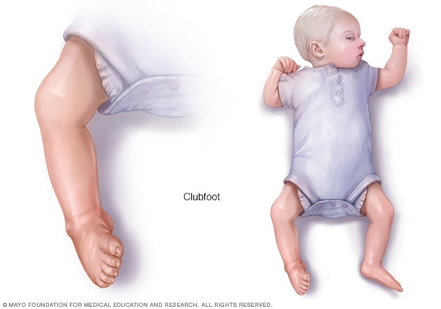
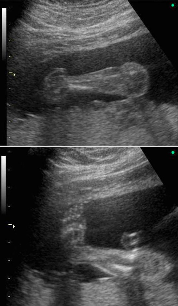
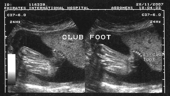
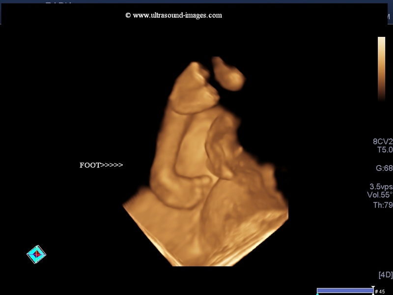
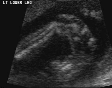
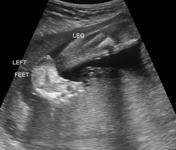
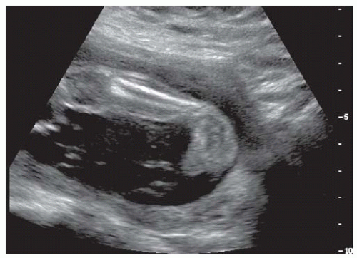
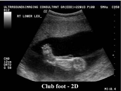
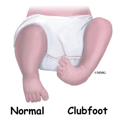
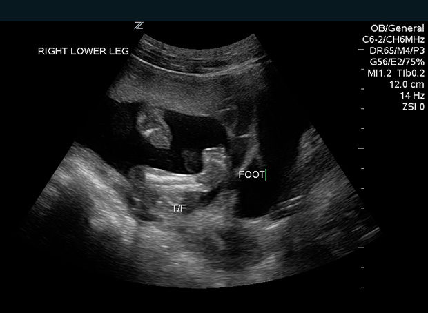
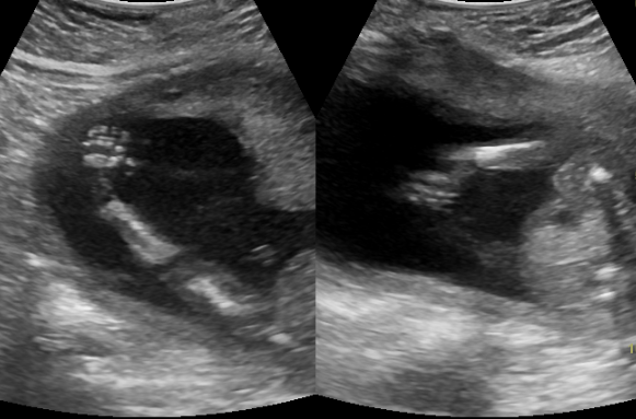
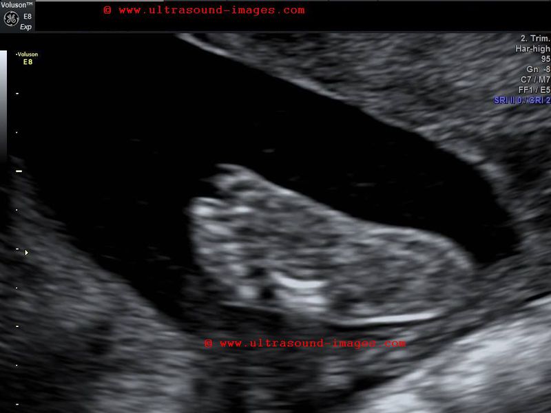
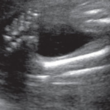
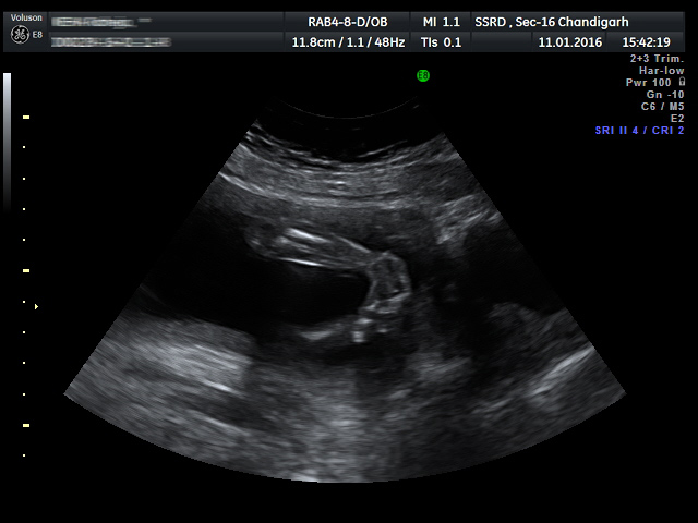
Club foot ultrasound images. A suspected diagnosis of clubfoot can be determined via prenatal ultrasound as early as 13 weeks, but it is typically discovered during an ultrasound around weeks gestation. Clubfoot, or talipes equinovarus, refers to a developmental deformity of the foot in which one or both feet are excessively plantar flexed, with the forefoot swung medially and the sole facing inward ().It is a common congenital malformation, typically discovered at the time of birth as an isolated anomaly in an otherwise normal neonate. In about half of the children with clubfoot, both feet are affected.
We just found out five days ago about the club foot and I picked up the imaging from the OB today to bring to the ortho specialist and of course I started looking up other ultrasound images to compare to and that's how I found your page. In most cases, the front of the foot is twisted downward and inward, the arch is increased, and the heel is turned inward. Clubfoot can also be diagnosed by a doctor immediately after a baby is born.
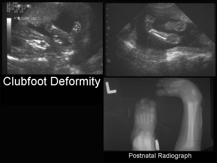
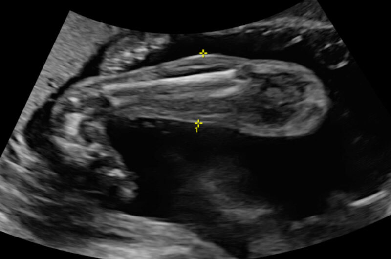
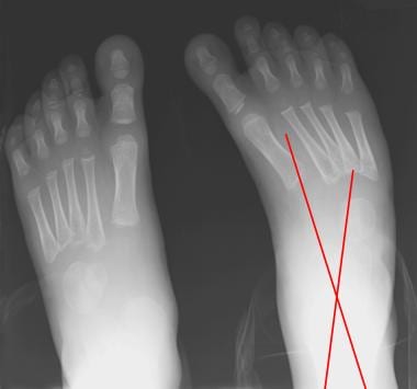
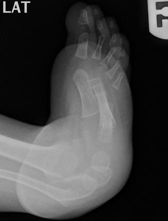
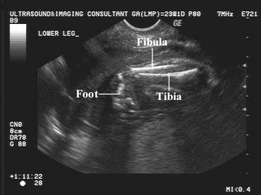
If soft tissue damage or tissue inflammation is suspected an MRI or diagnostic ultrasound may be indicated. Imaging the entire foot in the same plane as tibia/fibula is recognized as a simple hallmark in imaging for club foot. Clubfoot is a birth defect where one or both feet are rotated inward and downward.
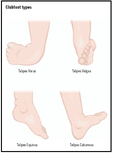
It’s easy to correct in most cases, so most children don’t have long-lasting. Musculoskeletal ultrasound (US) is a rapidly evolving technique that is gaining popularity for the evaluation and treatment of joint and soft tissue diseases. Talipes equinovarus (adduction of the forefoot, inversion of the heel and plantar flexion of the forefoot and ankle);.
We, therefore, deem that any gap measurements are, in fact, useless. FETAL FOOT These are ultrasound images of a 2nd trimester fetus, which show a neural tube defect (spina bifida) of the lower lumbar vertebrae. Foot ultrasound of plantar fibromatosis, plantar plate, tear, rupture, mortons neuroma, mulders click, metatarsalgia, effusion, foreign body GooGhywoiu99t543j0s7543uw1.
A 3D ultrasound image of the total embryo was taken. Imaging such as ultrasound, CT, and MRI are considered, conventional radiographs are initially obtained in a variety of acquired and congenital disorders of the foot. Ultrasound of the Foot and Ankle is a step-by-step introduction on how to use ultrasound successfully to diagnose and treat conditions of the foot and ankle.
This report will show any problems with your pelvic organs, blood vessels, or unborn baby. The penis on the male fetus appears as what is commonly called the "turtle" sign, which resembles like a turtle poking its head out of its shell. There are several things that can cause club foot like position, t18, and ehlers-danlos syndrome or it could be just a bad ultrasound image.
Five cases of congenital clubfoot diagnosed prenatally by ultrasound are reported. It can also be detected before birth by ultrasound, especially if both feet are. Keywords clubfoot, talipes equinovarus, talipes, clubfeet Introduction Talipes equinovarus (clubfoot) is an abnormality of the foot.
Clubfoot, or talipes equinovarus, is a congenital deformity consisting of hindfoot equinus, hindfoot varus, and forefoot varus.The deformity was described as early as the time of Hippocrates. As the diagnosis of a congenital clubfoot is based on the subjective assessment of the ultrasound images, there is, especially at the end of the first trimester, a need for a measurement tool for objective documentation of the foot position. Doctors use the term "clubfoot" to describe a range of foot abnormalities usually present at birth (congenital).
Ultrasound can give us two-dimensional, and in some applications three-dimensional, images of structures and organs in virtually any part of the body. Browse 7,4 ultrasound stock photos and images available, or search for ultrasound baby or pregnancy ultrasound to find more great stock photos and pictures. Abstract In this chapter, we review the diagnosis and underlying potential causes for isolated clubfoot (Talipes Equinovarus).
The results documented a stage of "physiologic club foot" characterized by a medial deviation and plantigrade orientation of talar neck and. Clubfoot doesn’t cause pain, but if it’s not treated, it can make it hard for a child to walk without a limp. The advantages of sonography include its unsurpassed depiction of normal.
Although measurements have been given for the exact tenotomy gap and tendon widths in static ultrasound images, no mention was made of the position of the foot, which we have found has a large impact on gap measurements 6–9 (Figs. At 22 weeks pregnant, I had a Level II ultrasound at a perinatalogist's practice to confirm the diagnosis and also to check for any other issues (sometimes, clubfoot can be an indication of a larger problem such as spina bifida). Magnetic resonance provides a more uniform and reproducible image for long-term follow-up studies.
Schwartz, DPM, the book thoroughly covers both diagnostic and interventional methods. Select Category Abdomen and retroperitoneum. If instability is suspected a stress image can be taken to determine ligamentous laxity.
Dec 2, 17 - Explore randapaige's board "club foot baby!!", followed by 842 people on Pinterest. Most diseases and conditions are often a result of genetic or environmental factors. I haven't even explored the rest of your page but I surely will.
In addition to diagnostic uses, such as evaluating abnormalities in the abdomen, pelvis, and breast, ultrasounds are commonly used to guide needle and catheter placement in a variety of surgical. Most of the time, it is not associated with other problems. 1.1 Liver 1.2 Gallbladder and bile ducts 1.3 Pancreas 1.4 Spleen 1.5 Appendix 1.6 Gastrointestinal tract 1.7 Peritoneum mesentery and omentum 1.8 Various intra-abdominal tumors 1.9 Retroperitoneum and great vessels 1.10 Adrenal glands 1.11 Abdominal wall 1.12.
Ultrasound imaging can identify the sex of the baby by imaging the groin area. Even though in many cases, the causes of clubfoot is usually persistent with the position of the baby while he or she is in the womb of the mother which is called Postural Clubfoot. At birth, a doctor will examine your baby’s feet, arms, hands, hips and legs.
Club foot ultrasound the fetus with congenital club foot. Club foot is a relatively common finding during antenatal scan. Thankfully, everything else looked great and the clubfoot was the only anomaly the tech could see.
Using ultrasound to image the foot in rheumatoid arthritis:. Clubfoot, or talipes equinovarus (TEV), is commonly diagnosed on prenatal ultrasound. Thank you for writing this.
Many parents find out their child has clubfoot during a prenatal ultrasound months or weeks before their child is born. When we talk about clubfoot causes, it’s the same. Club Foot - Symptoms, Causes, Treatment, Surgery information and Pictures, images.Clubfoot hinders the development of the child especially when it is time for the child to start walking.
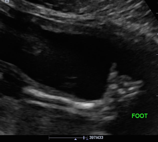
Talipes calcaneovalgus (dorsal flexion of the forefoot with the plantar surface. While nothing can be done before birth to solve the problem, knowing about the condition may give you time to learn more about clubfoot and get in touch with appropriate health experts, such as a pediatric orthopedic. This results in the dorsum of the foot being rotated medially with the ultrasound images showing the metatarsals and phalanges of the affected foot in the same view and same plane as the tibia and fibula of the lower leg.
This condition is found to be more common in children, especially the female ones. There is also splaying of the vertebral laminae and widening of the interpedicular distance in this region. E:FETAL CLUB FOOT F:.
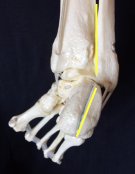
The term talipes is derived from a contraction of the Latin words for ankle, talus, and foot, pes.The term refers to the gait of severely affected patients, who walked on their ankles. Clubfoot is almost always diagnosed during a prenatal ultrasound—a technique that uses high-frequency sound waves to create images of babies in the womb. Prenatal ultrasound (US) findings in particular are described, along with accompanying images to augment the reader's understanding.
Without treatment, the foot remains deformed, and people walk on the sides of their feet. See more ideas about Club foot baby, Club foot, Baby feet. Causes of Clubfoot :.
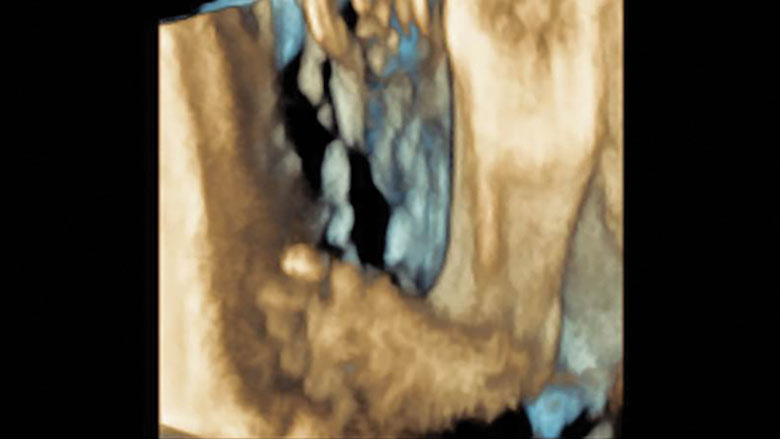
Three‐dimensional ultrasound allows precise alignment of orthogonal planes in which accurate measurements can be made and allows creation of rendered casts of the irregularly shaped mandibular bone. 1 Faculty of Health Sciences, University of Southampton, Building 45, University Road, Southampton, Hampshire, SO17 1BJ, UK. About 50 percent of children with clubfoot have it in both feet, a condition known as bilateral clubfoot.
Get information and make a plan. Around 10% of babies with clubfoot have another fetal condition. Deriving measurements from two‐dimensional and three‐dimensional images 6-8.
A clear understanding of normal US anatomy is required to prevent misdiagnosis and ensure optimal patient care. This study sought to visualize TEV and associated abnormalities on fetal magnetic resonance imaging (MRI. A radiologist will analyze the ultrasound images and send a report to your doctor.
A clubfoot is a congenital deformity in which the affected foot appears rotated internally at the ankle. Ultrasound Case 8- Ultrasound Images of an interdigital neuroma Ultrasound Case 9- Ultrasound Images of an interdigital neuroma The Ankle, Foot and Orthotic Centre’s Northcote Podiatrists can help you with all lower limb complaints, including Interdigital Neuromas. Typically, clubfoot affects both feet, though some babies are born with only one clubfoot.
Clubfoot, or talipes equinovarus, is a deformity in which the foot is excessively plantar flexed, with the forefoot bent medially and the sole facing inward.This usually results in the underdevelopment of the soft tissues on the medial side of the foot and calf and to various degrees of rigidity of the foot and calf. Doctors use a number of different imaging procedures to look at the structures within our feet and ankles. Hallux Valgus is a deformity where the great toe deviates laterally and the 1st metatarsal deviates medially to create a valgus angle.
Synovitis Introduction The small joints of the hands and feet play a central role in the diagnosis and classification of arthropathy. Diagnosing Clubfoot Doctors can see clubfoot on ultrasound images taken after about 4 months of pregnancy. 2 NIHR Musculoskeletal Biomedical Research Unit, University of.
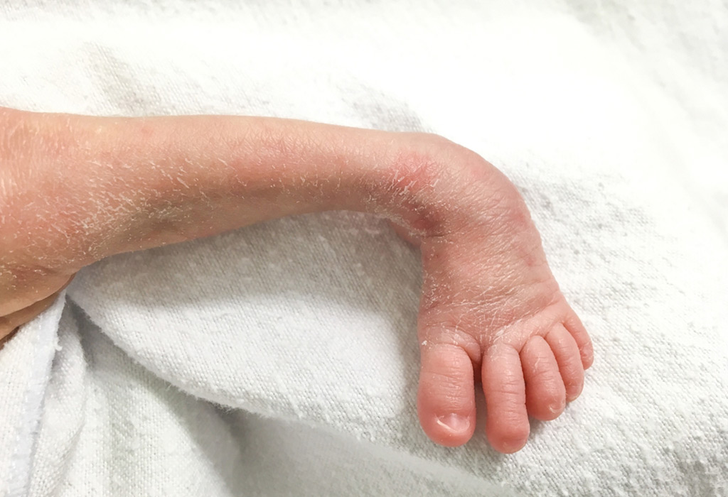
The severity of the clubfoot often cannot be determined until after delivery. Patient was born with deformity. Remember how precious your little one is and work through it together.
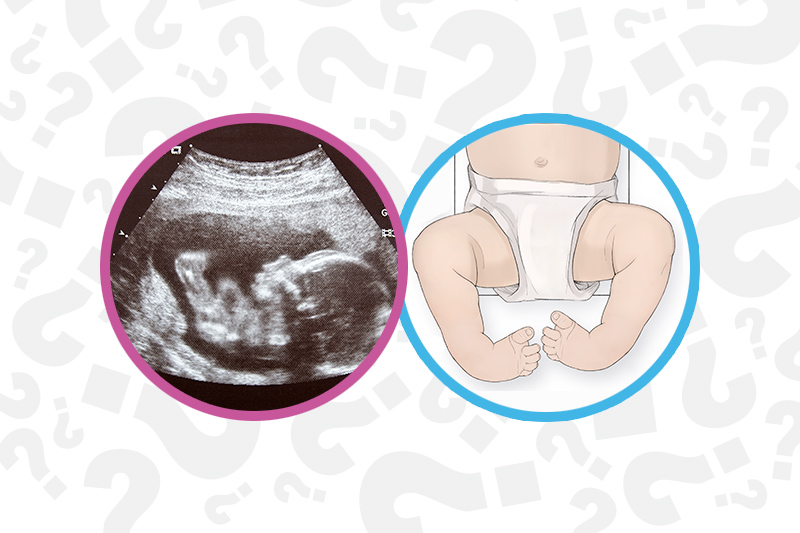
Authored by noted podiatric physician and educator, Nathan H. In clubfoot, the tendons that connect the leg muscles to the foot bones are short and tight, causing the foot to twist inward. This also results in the foot having a club like appearance and is called club foot or congenital talipes equinovarus.
One of the most commonly used of these procedures is foot and ankle ultrasound (also known as a sonogram), which uses high-frequency sound waves to form an image of the bones and other parts of the foot and ankle.This technique has been around for over fifty years, and it is useful for. It's possible to clearly see most cases of clubfoot before birth during a routine ultrasound exam in week of pregnancy. The affected foot and leg may be smaller in size compared to the other.
Explore {{searchView.params.phrase}} by color family {{familyColorButtonText(colorFamily.name)}}. The incidence of clubfoot may be higher within an affected family and may be associated with other structural anomalies or chromosomal abnormalities. Clubfoot, otherwise known as talipes equinovarus, is a deformity affecting the foot and the ankle wherein it is turned inward and downward.
Approximately 50% of cases of clubfoot affect both feet. Images were reviewed by the senior radiologist (D.H.P.) to determine the modality of limb abnormality diagnosis (transabdominal sonography versus transvaginal sonography), utility of 3‐dimensional (3D) sonography in aiding diagnosis of the limb abnormality, and the relevance of the limb abnormality in aiding the overall chromosomal or. 3-D ultrasound may narrow this gap.
Once the child is born, the condition is clearly visible. Ideally, treatment begins in the first month of a child’s life. An Explanation of Technique and Terms Although some of this information is repeated in the case scenarios to follow, it is helpful to begin by discussing the.

Figure 2 From Congenital Talipes Equinovarus A Case Report Of Bilateral Clubfoot In Three Homozygous Preterm Infants Semantic Scholar
Clubfoot Symptoms And Causes Mayo Clinic

Clubfoot Or Congenital Talipes Equinovarus Ctev Miracles Mediclinic
Club Foot Ultrasound Images のギャラリー
Club Foot Radiology Case Radiopaedia Org
Dynamic Real Time Ultrasound Of The Clubfoot And Ankle Joint Sonoskills

2d 3d 4d Ultrasound Of The Fetal Face In Genetic Syndromes Radiology Key

Club Foot Jax S Journey

Club Foot

My Journey With Baby S Positional Clubfoot Part 1 Baby Gizmo

Congenital Fetal Anomalies And The Role Of Prenatal Ultrasound Intechopen

Prenatal Ultrasound Diagnosis Of Club Foot The Bone Joint Journal
Congenital Talipes Equinovarus Radiology Reference Article Radiopaedia Org
Embryo With Xyy Syndrome Presenting With Clubfoot A Case Report Cases Journal Full Text
Www Pedrad Org Portals 5 Events 13 Rypenshandsandfeet Pdf
Clubfoot Deformity Talipes Equinovarus
A Gallery Of High Resolution Ultrasound Color Doppler 3d Images Fetal Spine
Introduction To Clubfoot Physiopedia

Prenatal Ultrasound Diagnosis Of Club Foot The Bone Joint Journal
A Gallery Of High Resolution Ultrasound Color Doppler 3d Images Fetal Face And Neck

Skeleton Diagnosis Of Fetal Abnormalities The 18 23 Weeks Scan
When Your Baby Has Clubfoot Answers For Expecting Parents Boston Children S Discoveries
When Your Baby Has Clubfoot Answers For Expecting Parents Boston Children S Discoveries

Clubfoot Deformity Talipes Equinovarus
Antenatal 3d Usg In Unilateral Club Foot A Rare Anomaly Insight Medical Publishing

Protected Blog Log In Ultrasound Club Foot How To Apologize

Ultrasound Video Showing Flat Foot Deformity Rocker Bottom Feet Youtube

Ultrasound Video Showing Club Foot Fetal Anomaly Scan Youtube

Congenital Talipes Equinovarus Radiology Case Radiopaedia Org
Q Tbn 3aand9gcrhcde8f5w0l798munyrho 8myptyhufww5xdkf93yfpwmrn8wm Usqp Cau

404 Not Found Ultrasound Sonography Ultrasound Sonography

Club Foot Ultrasound Youtube

Prenatal Clubfoot Increases The Risk For Clinically Significant Chromosomal Microarray Results Analysis Of 269 Singleton Pregnancies Sciencedirect

Pdf Prenatal Sonographic Diagnosis Of Talipes Equinovarus Clubfoot In A Non Selected Population Of Northwest Of Iran

The Catholic Working Mother June 13

Pdf Prenatal Ultrasound Diagnosis Of Club Foot Outcome And Recommendations For Counselling And Follow Up Semantic Scholar

Club Foot In Ultrasound Babycenter

Club Foot
Clubfoot Deformity Talipes Equinovarus
Congenital Talipes Equinovarus Radiology Reference Article Radiopaedia Org
Obgyn Onlinelibrary Wiley Com Doi Pdf 10 1046 J 1469 0705 1998 1103 X

Reviews Chews How Tos Penelope S Clubfoot Journey So Far

My Baby Has A Club Foot Babycenter

Club Foot

Club Foot Nidirect
Value Of The Fetal Plantar Shape In Prenatal Diagnosis Of Talipes Equinovarus Liao 12 Journal Of Ultrasound In Medicine Wiley Online Library
The Foot Musculoskeletal Key
Clubfoot Symptoms Stages Definition Description Demographics Causes And Symptoms Diagnosis
Q Tbn 3aand9gcq8z1guuraslaxxswsdzncaggejwuzgp7mw Bpx1jez9lbxnwuu Usqp Cau

Fetal Skeletal System Diagnostic Medical Sonography Medical Ultrasound Ultrasound

Clubfoot Versus Positional Foot Deformities On Prenatal Ultrasound Imaging Brasseur Daudruy Journal Of Ultrasound In Medicine Wiley Online Library

My 1 In 1 000 Triad Moms On Main Greensboro Winston Burlington High Point

The Clubfoot Chronicles The Saga Begins

Prenatal Ultrasound Of Case 2 At 34 Gestational Weeks Shows A A Download Scientific Diagram

Club Foot
Www Ajog Org Article S0002 9378 19 3 Pdf

Fetal Clubfoot Lurie Children S
Q Tbn 3aand9gcs1kvzryv9liujzesyxzpihxt3hxi49fewapmni3e 8uiol3w4o Usqp Cau

Prenatal Diagnosis Of Clubfoot A Review Of Current Available Methodology Topic Of Research Paper In Clinical Medicine Download Scholarly Article Pdf And Read For Free On Cyberleninka Open Science Hub

Antenatal 3d Usg In Unilateral Club Foot A Rare Anomaly Semantic Scholar

Wendy Davis Would Be Okay If Club Footed Babies Like My Son Are Aborted Lifenews Com

Fetus General Normal Fetal Anatomy Ultrasound Services Service Provider From Ernakulam
Clubfoot Deformity Talipes Equinovarus
Tackling Talipes Early With A Team Approach Children S Hospital Of Philadelphia
Clubfoot Eorthopod Com

Congenital Fetal Anomalies And The Role Of Prenatal Ultrasound Intechopen
Club Foot

First Trimester Physiological Development Of The Fetal Foot Position Using Three Dimensional Ultrasound In Virtual Reality Bogers 19 Journal Of Obstetrics And Gynaecology Research Wiley Online Library

Club Foot By Scout Martin

Please Put My Mind At Ease March Babies Forums What To Expect

My Journey With Baby S Positional Clubfoot Part 1 Baby Gizmo

My Journey With Baby S Positional Clubfoot Part 1 Baby Gizmo

Fetal Clubfoot Ultrasound Services In Ernakulam Ultrascan Centre Id

Clubfoot Causes And Treatments
Club Foot Antenatal Ultrasound Image Radiopaedia Org

Three Dimensional 3d Ultrasound Studies A Bilateral Club Foot Download Scientific Diagram

Bilateral Congenital Talipes Equino Varus Deformity In Fetus Radiology Case Radiopaedia Org
A Gallery Of High Resolution Ultrasound Color Doppler 3d Images Fetal Face And Neck

Clubfoot Talipes Equinovarus Radiology Key
Clubfoot Deformity Talipes Equinovarus

Prenatal Ultrasound To Detect Fetal Anomalies American Academy Of Pediatrics

Correlations Between Physical And Ultrasound Findings In Congenital Clubfoot At Birth Sciencedirect
Q Tbn 3aand9gcq8z1guuraslaxxswsdzncaggejwuzgp7mw Bpx1jez9lbxnwuu Usqp Cau
Clubfoot Deformity Talipes Equinovarus

Clubfoot
Clubfoot Imaging Practice Essentials Radiography Computed Tomography

Foot Problems Pediatrics Clerkship The University Of Chicago
Ultrasound Images Of Fetal General
Clubfoot Congenital Talipes Equinovarus Pediatrics Orthobullets

Skeleton Diagnosis Of Fetal Abnormalities The 18 23 Weeks Scan
Tackling Talipes Early With A Team Approach Children S Hospital Of Philadelphia
Clubfoot Congenital Talipes Equinovarus Pediatrics Orthobullets
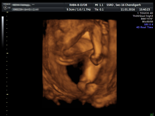
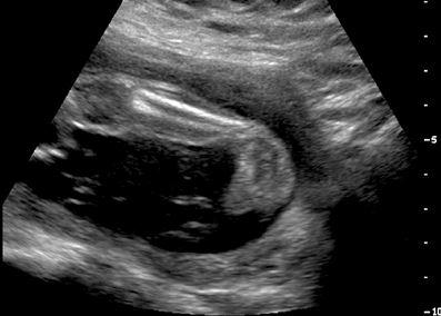
Ssrd Interesting Cases Fetal Clubfoot Ultrasound Image 3d Image

Club Foot Talipes Equinovarus Ankle Foot And Orthotic Centre

Congenital Talipes Equinovarus Radiology Reference Article Radiopaedia Org
Clubfoot Deformity Talipes Equinovarus
Club Foot Nhs

Ultrasound And X Rays For Foot Care Podiatry Associates P C

Clubfoot At 22 Second Weeks Gestation The Forefoot Arrows Is Download Scientific Diagram
Ssrd Interesting Cases Fetal Clubfoot Ultrasound Image 3d Image

Ultrasound Evaluation Of Clubfoot Correction During Ponseti
Club Foot In Infants Reasons Signs Remedies

Skeleton Diagnosis Of Fetal Abnormalities The 18 23 Weeks Scan

Club Foot Interactive Health

Club Foot Antenatal Ultrasound Radiology Case Radiopaedia Org


