Baby Ultrasound Club Foot
Another reason you may have an ultrasound at 16 weeks is to check the length of your cervix.

Baby ultrasound club foot. It really has no effects on your newborn. Clubfoot is diagnosed through ultrasound and can only be treated after birth with casting and/or reconstructive surgery. Signs of clubfoot include a short and/or tight Achilles tendon (heel cord) and a heel that is turned in.
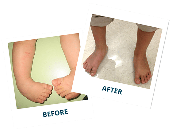
About half of children with clubfoot have it in both feet. It’s when a baby’s foot turns inward so that the bottom of the foot faces sideways or even up. However, it does need to be corrected, as it will not resolve on its own.
What You Need To Know When it comes to our babies, we are extremely protective and want to ensure the best for them. An orthopedic surgeon should be contacted to discuss what options for treatment are available. A questionnaire assessment indicated that 17 (65.4%) deemed that the explanation of the baby's condition was clear.
· Incidence ranging from about 0.1% in the newborn population to 0.4% when diagnosed antenatally by ultrasound (1). To determine the outcome of fetuses with clubfoot diagnosed by prenatal sonography. How is clubfoot diagnosed?.
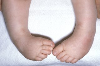
Clubfoot Clubfoot is a birth defect that causes a child’s foot to point inward instead of forward. Clubfoot can also be diagnosed by a doctor immediately after a baby is born. In clubfoot, the tendons that connect the leg muscles to the foot bones are short and tight, causing the foot to twist inward.
· with a reported incidence of 0.4 per 1000 live births in Chinese, · 1 to 5 per 1000 in whites, · 6 to 8 per 1000 in Polynesians (2). Treatment for clubfoot usually begins within a few weeks of a child’s birth. A positional clubfoot is flexible, rather than rigid, and can be positioned into a neutral position easily by hand.
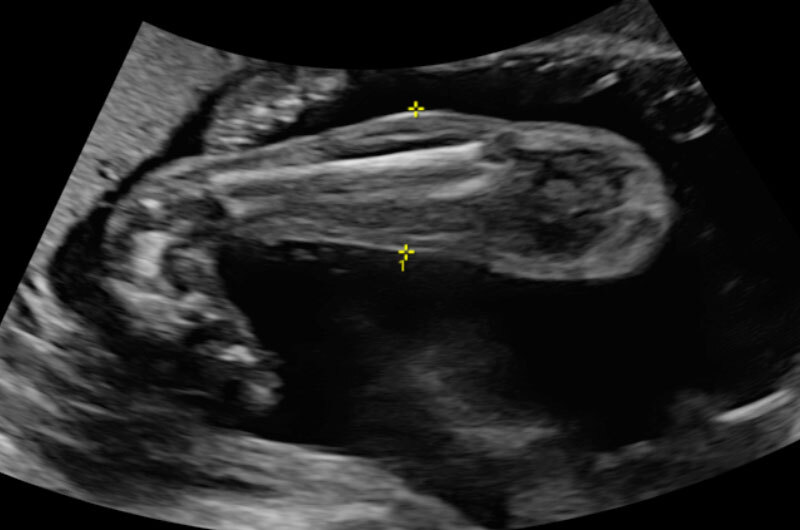
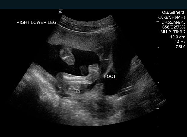
Without treatment, persons afflicted often appear to walk on their ankles, or on the sides of their feet. Published on Feb 01, 18 in In Utero Insights 2-D ultrasound of clubfoot Talipes (known as clubfoot) is one of the most common congenital foot and ankle problems, occurring in about one in 1,000 live births and in boys twice as often than in girls. How does clubfoot affect my baby?.
Prenatal ultrasound diagnosis of clubfoot is increasing. Clubfoot can range from mild to severe. If a mother responded that the diagnosis occurred by prenatal ultrasound, the clubfoot was determined to be prenatally diagnosed.
O35-Maternal care for known or suspected fetal abnormality and damage ICD-10-CM Diagnosis Code O35.8XX0. Club foot is usually diagnosed after a baby is born, although it may be spotted in pregnancy during the routine ultrasound scan carried out between 18 and 21 weeks. A clubfoot, or talipes equinovarus 1 (TEV), is a birth defect.
It can also be detected before birth by ultrasound, especially if both feet are. Then, he wore a brace for 6 months, then a special insole in his shoe for a while. About one out of every 1,000 babies is born with a foot that’s twisted.
About half of babies with clubfoot have it in both feet and approximately 1 to 4 babies in 1,000 are born with this condition. My son was born with severe club foot on the right back in 06. Clubfoot is usually diagnosed during a newborn exam, though it can sometimes be seen during a fetal ultrasound in the womb.While it can be very upsetting to learn that your baby has a deformity, the good news is that treatment for clubfoot is highly successful, especially when therapy starts right after birth (while your newborn’s bones, joints, and tendons are extremely.
Clubfoot is a birth defect where one or both feet are rotated inward and downward. Club foot is quite common, affecting about 1 baby in every 1,000 born in the UK. Of 103 patients with clubfoot diagnosed at birth, 26 (25.2%) positive prenatal scans were identified with the earliest diagnosis being made at 15 weeks.
Most of the time, a baby’s clubfoot is diagnosed during a prenatal ultrasound before they are born. O30-O48 Maternal care related to the fetus and amniotic cavity and possible delivery problems ;. The front half of an affected foot turns inward and the heel points down.
Once the condition has been detected, a targeted ultrasound will be performed to rule out the presence of associated anomalies. Anyone else experience this or been told their baby is club foot and then is born without club foot?. About 10 percent of clubfeet can be diagnosed as early as 13 weeks into pregnancy.
We just had our 19 week scan and my husband and I were told our baby boy could have club foot. Clubfoot is a common birth defect that causes the front of the foot to turn inward and the heel to point down. If your child has clubfoot, it will make it harder to walk normally, so doctors generally recommend treating it soon after birth.
It occurs twice as often in males than in females. While the condition isn’t painful for babies, if left untreated it can lead to limb deformities and problems walking later in life. Although clubfoot is diagnosed at birth, many cases are first detected during a prenatal ultrasound.
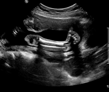
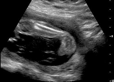
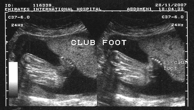
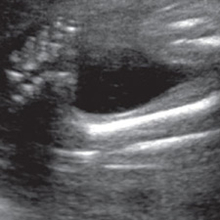
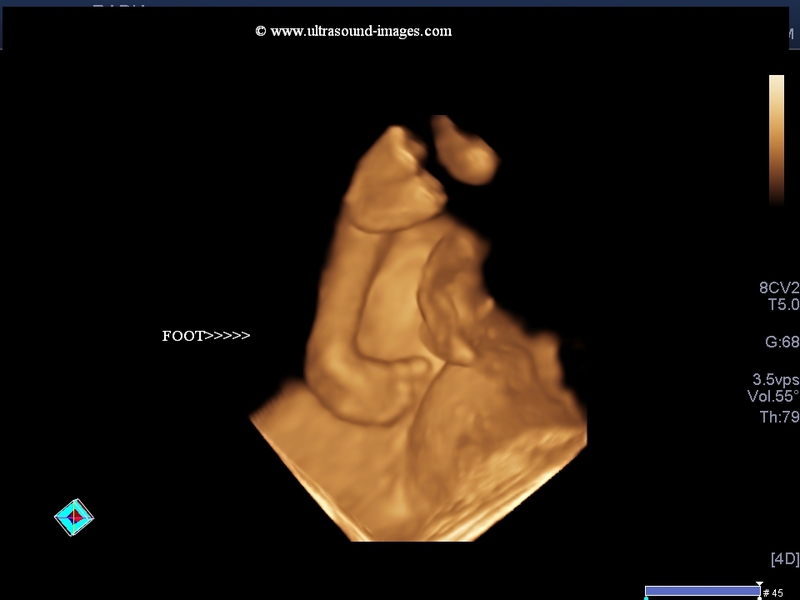
This video shows Fetal Club foot (CTEV) and Soft tissue edema. A baby with club foot has a foot that resembles the end of a golf club (hence its name). Fetal clubfoot can be diagnosed by ultrasound (sonogram) examination before birth.
Clubfoot also can be discovered in utero (while the baby is still in the mother’s womb) during an ultrasound. Clubfoot, also known as talipes equinovarus (TEV), is a common foot abnormality, in which the foot points downward and inward. ICD-10-CM Codes › O00-O9A Pregnancy, childbirth and the puerperium ;.
Occasionally, the doctor may request X-rays to fully understand how severe the clubfoot is, but usually X-rays are not necessary. Preterm Labor Risk Assessment. The heel points down and the front half of the foot turns in.
Your baby's foot bones, muscles, tendons, and blood vessels may also be affected. Both feet are affected in about half of these babies. The prenatal detection of intrauterine growth restriction, hypospadias and clubfoot should raise the suspicion of WHS.
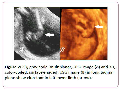
Club foot deformity can usually be identified on prenatal ultrasound exam. High-resolution banding and molecular analysis can help to confirm the diagnosis. Or, it might have an odd shape and point in the wrong direction, so.
Most of the time, it is not associated with other problems. Clubfoot is a common birth defect. Club foot might be diagnosed on an ultrasound examination before the baby is born.
One or both feet may be affected. While nothing can be done for the infant before birth, parents can learn about the condition. As many as four children out of every 1,000 are born with clubfoot.
Boys have clubfeet nearly twice as often as girls. Treatment of Club Foot. Get information and make a plan.
There are several things that can cause club foot like position, t18, and ehlers-danlos syndrome or it could be just a bad ultrasound image. It may curl sideways with the toes bent at a funny angle. · The incidence of clubfoot varies with ethnicity:.
In about half of the children with clubfoot, both feet are affected. Congenital talipes equinovarus, often also known as ‘club-foot’, is a common developmental disorder of the lower limb. Subtle abnormalities on ultrasound may suggest a chromosomal problem.
It is a common birth defect, occurring in about one in every 1,000 live births. We identified all fetuses scanned at our institution from May 19 to May 02 in whom clubfoot was suspected or diagnosed on prenatal sonography. Without treatment, the foot remains deformed, and people walk on the sides of their feet.
Unilateral or bilateral clubfoot, gestational. Visual identification of club foot is all that is needed for diagnosis. The condition, also known as talipes equinovarus, is fairly common.
Typically, clubfoot affects both feet, though some babies are born with only one clubfoot. Clubfoot is the most common foot deformity present at birth, occurring in approximately 1 in every 1,000 births. An ultrasound is a type of imaging used to look at babies in the womb.
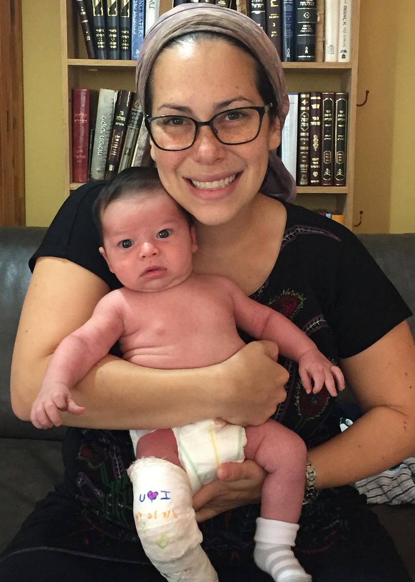
My son had to have casts on his foot for a year, before surgery to lengthen his achilles tendon at 1 year old. Clubfoot actually describes an array of foot deformities that cause your newborn baby's feet to be twisted, pointing down and inward. Clubfoot is a congenital foot deformity that affects a child’s bones, muscles, tendons, and blood vessels.
Some subtle cases may be missed on ultrasound but are easily diagnosed after birth. It is routine for a woman to have an ultrasound during her pregnancy to confirm her baby’s growth and development. From maternal and neonatal medical records, we collected the following information when available:.
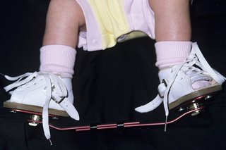
Clubfoot is a condition in which the tendons and ligaments in a baby’s foot and ankle are abnormally short and tight. However, diagnosis based on ultrasound alone produces a % false positive rate. Treatment There are two treatments currently used to treat club foot – the Ponseti Method and surgery.
Collin May of the Lower Extremity Program in Boston Children’s Orthopedic Center answers questions about how meeting with an orthopedic specialist can ease expecting parents’ fears about a. The condition is normally identified after birth, but doctors can also tell if an unborn baby has. A relatively common birth defect and typically an isolated problem for an otherwise healthy baby, clubfoot refers to a range of foot abnormalities in which the one or both of the baby’s feet is twisted out of shape or position.
Diagnosis The condition can be diagnosed in-utero via ultrasound or at birth. In severe cases, the foot is turned so far that the bottom faces sideways or up rather than down. This is typically done if you delivered a preterm baby (before 37 weeks) in a prior pregnancy.Unlike the typical prenatal ultrasound, this is a transvaginal ultrasound and is done only to measure your cervix.
Our Baby Baby Boy Baby Club Calf Muscles Baby Health Change My Life Ultrasound Medical Conditions Family History Clubfoot In Babies:. Responses included prenatal ultrasound, examination at birth, examination at follow-up pediatrician visit, or other. Your maternal-fetal medicine specialist will do a more extensive ultrasound to confirm the diagnosis and look for any other anomalies your child may have.
Approximately 10% of all clubfeet can be diagnosed by 13 weeks gestation, and about 80% can be diagnosed by 24 weeks gestation. Clubfoot is almost always diagnosed during a prenatal ultrasound—a technique that uses high-frequency sound waves to create images of babies in the womb. Start researching ortopedic specialists that have lots of experience with club foot.
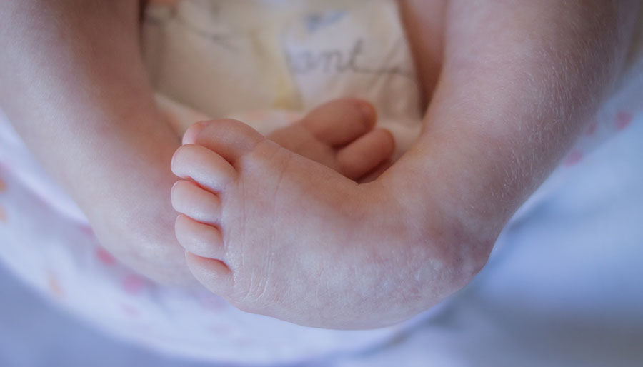
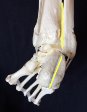
Clubfoot is not painful for your baby. Clubfoot, also known as talipes equinovarus, is a condition where a baby’s foot is twisted inward to the point where the bottom of the foot faces sideways and in some cases upward. The shortened tendons cause the feet to turn inward and downward.
Most commonly, a doctor recognizes clubfoot soon after birth just from looking at the shape and positioning of the newborn's foot. A positional clubfoot results from the baby's position in the uterus, and is often associated with a restrictive uterine environment (oligohydramnios, uterine anomalies). In my week ultrasound they said the baby might have unilateral clubfoot,in my 24 week ultrasound they confirmed it as bilateral clubfoot,in my 27 week ultrasound they said the left foot looks ok in one angle and they are not sure about the left foot and the right is clubbed,if it is a club feet it should be clubbed in all the angles right,do any of you have come across my situation,it's being so hard for me to push the days,i am depressed.
Like about half of children with the condition, the ultrasound showed that Cheryl and Sue’s baby would have clubfeet on both sides. The affected foot and leg may be smaller in size compared to the other. Surgery usually is not needed to correct club foot.
Your doctor can often detect clubfoot by ultrasound in the second trimester. Club foot is usually diagnosed after a baby is born, although it may be spotted in pregnancy during the routine ultrasound scan carried out between 18 and 21 weeks. Standard cytogenetics cannot always demonstrate a microdeletion.
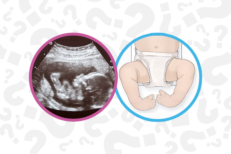
With prenatal ultrasound, parents often learn about clubfoot weeks or months before their child’s arrival. Clubfoot is a fairly common birth defect and is usually an isolated problem for an otherwise healthy newborn. Clubfoot is a birth defect that causes your baby's foot to point down and be turned inward.
It could just be the way his feet are, i was told with my daughter that she had club foot they did a second ultrasound to confirm itbut then my daughter was born perfectly normal, no club foot. The Achilles tendon (tissue that connects the heel to the muscles of the lower leg) is very tight, and calf muscles are smaller than normal. Club foot can’t be treated before birth, but picking up the problem during pregnancy means you can talk to doctors and find out what to expect after your baby is born.
Clubfoot (also called talipes equinovarus) is a birth defect of the foot. Mothers were then asked how the diagnosis of clubfoot first made. The foot is twisted in (inverted) and down.
In most cases, the foot appears to be pointing downwards and inwards. By 24 weeks, about 80 percent of clubfeet can be diagnosed, and this number steadily increases until birth. Approximately 50% of cases of clubfoot affect both feet.
This happens because the tissues that connect muscles to bone (called tendons) in your baby’s leg and foot are shorter than normal. Ultrasound Approximately 10-% of individuals with prenatally diagnosed clubfoot may have a normal foot or positional foot deformity requiring minimal treatment. The foot and ankle did not develop normally in utero.
We will have another ultrasound to confirm and treatment plan if needed. Club foot present as an in-turn of one foot or both feet. When a child has clubfoot, their feet and legs have all the same bones, tendons and muscles as a healthy child, but they are positioned incorrectly.

Learning About Club Foot
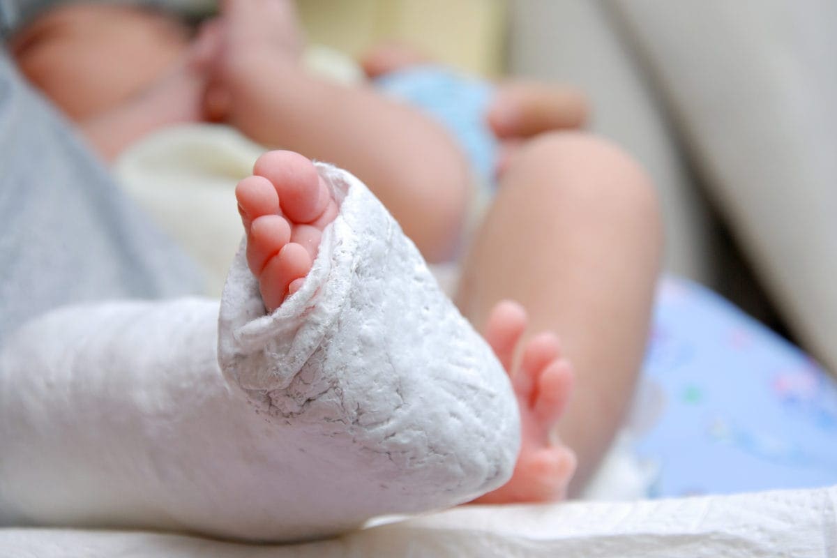
What Is Clubfoot Symptoms And Treatment Familydoctor Org

Do Not Stare Yes My Infant Has Braces On His Feet Pittsburgh Mom Collective
Baby Ultrasound Club Foot のギャラリー

Foot Problems Pediatrics Clerkship The University Of Chicago

A Step In The Right Direction Treating Clubfoot Sans Surgery Health Beat Spectrum Health

Clubfoot Deformity Talipes Equinovarus

2d 3d 4d Ultrasound Of The Fetal Face In Genetic Syndromes Radiology Key

My Journey With Baby S Positional Clubfoot Part 1 Baby Gizmo
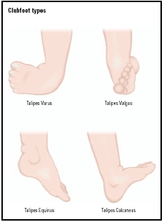
Clubfoot Symptoms Stages Definition Description Demographics Causes And Symptoms Diagnosis

Clubfoot Deformity Talipes Equinovarus

Clubfoot Wikipedia
The Catholic Working Mother June 13

Congenital Talipes Equinovarus Radiology Reference Article Radiopaedia Org

Fetal Clubfoot Lurie Children S

Skeleton Diagnosis Of Fetal Abnormalities The 18 23 Weeks Scan

Onelittlefoot Baby S Clubfoot Page 2

Skeleton Diagnosis Of Fetal Abnormalities The 18 23 Weeks Scan

Congenital Talipes Equinovarus Radiology Case Radiopaedia Org

Skeleton Diagnosis Of Fetal Abnormalities The 18 23 Weeks Scan
The Clubfoot Chronicles The Saga Begins

Fetal Skeletal System Diagnostic Medical Sonography Medical Ultrasound Ultrasound

Clubfoot Orthopaedia

Club Foot In Infants Reasons Signs Remedies

3d 4d Ultrasound Sydney Ultrasound Care
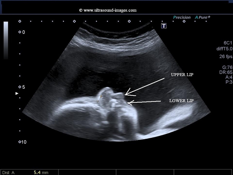
A Gallery Of High Resolution Ultrasound Color Doppler 3d Images Fetal Face And Neck
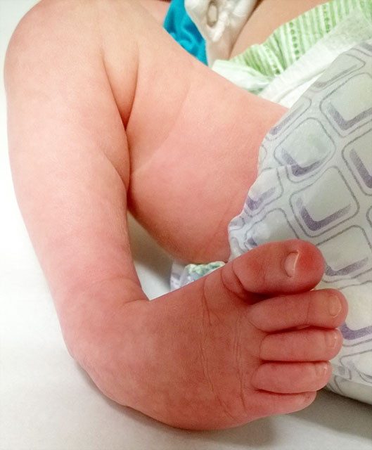
Clubfoot Johns Hopkins Medicine

Club Foot In Infants Reasons Signs Remedies
World Clubfoot Day Collin S Story

Trisomy 18 Clenched Fist Club Foot Html

Club Foot Talipes Equinovarus Ankle Foot And Orthotic Centre
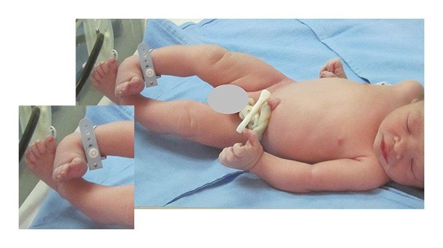
This Dad Is Glad He Got A Second Opinion On His Son S Clubfoot

Talipes Babycentre Uk

The Clubfoot Chronicles The Saga Begins

Trisomy 18 Clenched Fist Club Foot Html
Q Tbn 3aand9gcq8z1guuraslaxxswsdzncaggejwuzgp7mw Bpx1jez9lbxnwuu Usqp Cau

Club Foot In Ultrasound Babycenter

My Journey With Baby S Positional Clubfoot Part 1 Baby Gizmo
Club Foot

Amazing Results In Clubfoot Treatment For Young Riwan Steps
The Clubfoot Chronicles The Saga Begins
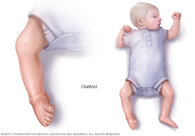
Clubfoot Symptoms And Causes Mayo Clinic

My Journey With Baby S Positional Clubfoot Part 1 Baby Gizmo
June 3rd World Clubfoot Day
Frontiers Developing A Three Dimensional 3d Assessment Method For Clubfoot A Study Protocol Physiology
Congenital Talipes Equinovarus Radiology Reference Article Radiopaedia Org
Clubfoot Congenital Talipes Equinovarus Pediatrics Orthobullets

The Clubfoot Chronicles The Saga Begins
When Your Baby Has Clubfoot Answers For Expecting Parents Boston Children S Discoveries

Club Foot Antenatal Ultrasound Radiology Case Radiopaedia Org

Please Put My Mind At Ease March Babies Forums What To Expect
Q Tbn 3aand9gcq8z1guuraslaxxswsdzncaggejwuzgp7mw Bpx1jez9lbxnwuu Usqp Cau

Clubfoot Deformity Talipes Equinovarus

Ultrasound Club Foot December 19 Babies Forums What To Expect
Clubfoot Orthoinfo os

Clubfoot Healthdirect

Raising A Newborn With Casts Kayla Vanaman

Clubfoot Deformity Talipes Equinovarus
Clubfoot A Mother S Story
Club Foot
Club Foot Nhs
Tackling Talipes Early With A Team Approach Children S Hospital Of Philadelphia

Clubfoot Treatment Bilateral Club Feet Foot Pain

Clubfoot In Newborns Paedicare Paediatricians
Club Foot Nhs

Club Foot Nidirect

My Baby Has A Club Foot Babycenter

Defying The Odds A Clubfoot Story News And Events Shriners Hospitals For Children Salt Lake City

Clubfoot For Parents Nemours Kidshealth
A Gallery Of High Resolution Ultrasound Color Doppler 3d Images Fetal Face And Neck
Overcoming Clubfoot One Mom S Story Parents

A Peachtree City Life An Update On Our Baby Girl Clubfoot

About Clubfoot

So You Re Expecting A Clubfoot Baby Thoughts Tips And Resources Cartwheeling Down The Aisle

Clubfoot Or Congenital Talipes Equinovarus Ctev Miracles Mediclinic
Marlowe S Clubfoot Journey How One Mom Went From Devastated To Reassured Children S Wisconsin

Club Foot

How Clubfoot Treatment Can Be Fun Kayla Vanaman

First Trimester Physiological Development Of The Fetal Foot Position Using Three Dimensional Ultrasound In Virtual Reality Bogers 19 Journal Of Obstetrics And Gynaecology Research Wiley Online Library

Protected Blog Log In Ultrasound How To Apologize Club Foot

A Step In The Right Direction Treating Clubfoot Sans Surgery Health Beat Spectrum Health
A Gallery Of High Resolution Ultrasound Color Doppler 3d Images Fetal Spine

How Parents And The Internet Transformed Clubfoot Treatment Shots Health News Npr
The Parents Guide To Clubfoot Miller Betsy Amazon Com Books

Club Foot
The Clubfoot Chronicles The Saga Begins
Q Tbn 3aand9gcrhcde8f5w0l798munyrho 8myptyhufww5xdkf93yfpwmrn8wm Usqp Cau
When Your Baby Has Clubfoot Answers For Expecting Parents Boston Children S Discoveries
Antenatal 3d Usg In Unilateral Club Foot A Rare Anomaly Insight Medical Publishing

Clubfoot Mendelson Kornblum Orthopedic Spine Specialists

Correlations Between Physical And Ultrasound Findings In Congenital Clubfoot At Birth Sciencedirect

Club Foot

Meet Our Focus For March Caius Club Foot Baby Club Foot Feet Treatment

Clubfoot Clubfoot Also Known As Pediatric Interesting Cases And Mcqs Facebook

Overcoming Clubfoot One Mom S Story Parents
Tackling Talipes Early With A Team Approach Children S Hospital Of Philadelphia
A Gallery Of High Resolution Ultrasound Color Doppler 3d Images Fetal Face And Neck
Introduction To Clubfoot Physiopedia

Predicting Recurrence After Clubfoot Treatment Lower Extremity Review Magazine

Clubfoot Deformity Talipes Equinovarus

Club Foot Shoes Baby Club Foot Club Foot Baby Baby Club

Clubfoot Causes And Treatments
Clubfoot Doctor Ladera Ranch Southern California Foot Ankle Specialists
Q Tbn 3aand9gcqxbbkoa0w3 Ib Elg 6rlmedprdorbjhrl4pycgcnakcuehefi Usqp Cau

Club Foot


